FAQ: Fibrolamellar

Frequently Asked Questions About Fibrolamellar Hepatocellular Carcinoma
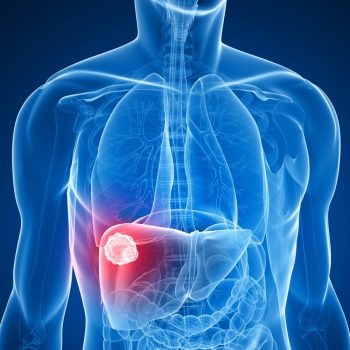
Fibrolamellar carcinoma (FLC or FLHCC) is a rare form of liver
cancer that mainly affects young adults. It is characterized by laminated
fibrous layers (“lamellae”) interspersed between tumor cells when
viewed under a microscope. Also known as fibrolamellar hepatocellular
carcinoma, it differs from the more common hepatocellular carcinoma (HCC) in
many factors including: its causes, its lack of known risk factors, markers in
the blood, response to drugs, and the age distribution of its patients – young individuals
with a normal, otherwise healthy liver (Lalazar and Simon, 2018; O’Neill et al., 2021;
Torbenson, 2012). The liver has a number of different cell
types including hepatocytes (which make up 80% of the mass of the liver and
carry out many of the classic functions we attribute to the liver),
cholangiocytes (which help form the ducts that run through the liver) and the
hepatic (meaning liver) stellate cells (which contribute many support functions
including immune responses, formation of blood vessels).
Tumors have been characterized by the organ they appear in (such as liver cancer) or the cells they appear in, thus the terms hepatocellular carcinoma, for a tumor in the hepatocytes or cholangiocarcinoma, for a tumor in a cholangiocyte (for more information on cholangiocarcinoma ). However, in the past ten years there has been a move toward “Precision Medicine” (House, 2015), characterizing a tumor based on the changes in the cells that drive the cancer. For example, the molecular change that drives fibrolamellar carcinoma is mainly found in hepatocytes, but has also been found in a few dozen cholangiocytes. Thus, instead of exclusively calling it a “hepatocellular” or “cholangiocarcinoma”, there is a shift from calling it “Fibrolamellar hepatocellular carcinoma” to the shortened “Fibrolamellar carcinoma.”
FLC has been traditionally viewed as a subvariant of hepatocellular carcinoma (HCC). However, there are a number of feature that distinguish the two and they are being viewed as distinct entities (Lalazar and Simon, 2018; O’Neill et al., 2021; Torbenson, 2012).
Patient population: FLC typically occurs in young patients, including children, adolescents and young adults, as young as 1 and usually under forty years of age with a median age of 22 at diagnosis. While HCC is also be found in adolescents, it is most commonly found in adults.
Cause: The exact cause of HCC is not clearly resolved. This is, in part, because HCC is a term that covers many different tumors that occur in the hepatocytes of the liver. HCC is often associated with damage, usually chronic damage, to the liver. This liver inflammation and cirrhosis (scaring of liver tissue) can be the result of chronic viral infections, alcohol overuse, or non-alcoholic fatty liver disease. However, there are patients who have HCC in the absence of any obvious damage to the liver. On the other hand, FLC arises in patients without liver inflammation, and their liver is usually otherwise healthy. FLC is caused by a specific mutation in a single liver cell (usually in a hepatocyte, but occasionally a cholangiocytes), leading to changes in the structure and function of the cell, which multiplies with the mass of mutated cells growing into a large tumor.
Structure of the tumor: Altered FLC tumor cells are different from those of HCC, and the larger tumors usually, but not always look different. FLC tumor tissue often has bands of fibers running in between the cells , giving fibrolamellar carcinoma its name . However, this different is not always so clear, and pathologists sometimes do not agree on a diagnosis based solely on how the tissue looks (Malouf et al., 2009).
Response to therapeutics: HCC and FLC respond differently to therapeutics. They are two very different diseases. One of the first major successes in treatment of HCC came from the use of sorafenib. In contract, there is no evidence to favor the use of sorafenib in FLC. So far, no system therapeutics have demonstrated sufficient efficacy against FLC to warrant approval by the FDA. Thus, surgical resection, when possible, is the mainstay of therapy. There is some evidence that surgical resection, even if it cannot completely remove the tumor, may extend survival (Berkovitz et al., 2022).
Our body’s cells share the same DNA, (arranged in chains known as “chromosomes”), which are large molecules containing instructions for cell functions. However, only specific parts of the DNA are relevant to each cell type and can vary even within the same cell. “Genes” are portions of DNA that serve as codes for specific actions in each cell. These codes are then translated into “proteins”, which are molecules that carry out various tasks in the different cells.
FLC is caused by a problem with a protein called protein kinase A (PKA). A kinase is a protein that can modify other molecules by adding to them a particular molecular group called a phosphate. This modification can turn on some molecules and turn off others. Many cancers are the result of either mutations in a kinase or overexpressing a kinase. In most FLC patients, a specific genetic change (“mutation”) occurs, where a segment of DNA on the 19th chain (chromosome 19) is somehow lost (a DNA “deletion”), with the 2 genes standing at each of its ends, DNAJB1 and PRKACA, fusing together. This is abbreviated in scientific text as DNAJB1::PRKACA. PRKACA is a gene that codes the active component of PKA. This results in the production of a fusion protein DNAJB1::PRKACA, instead of the normal PKA that exists in normal cells. It is not yet resolved how the “fusion” alters the cells. There is evidenced that fusion of a different protein, ATP1B1 (instead of DNAJB1) to PRKACA can also produce FLC (Singhi et al., 2020). Also, there are examples of patients who have FLC only by have increased activity of PKA, but without the DNAJB1 fusion (Graham et al., 2018). Thus, central to all of these cases of FLC is a disruption in the signaling of the protein kinase A pathway.
It has been estimated that 200 new cases are diagnosed worldwide each year (Lalazar and Simon, 2018; O’Neill et al., 2021). However, a recent study in the USA, analyzing electronic medical records in a local area and extrapolating the incidence to US nationwide populations, suggests it may be at least five to eight times more common (Zack et al., 2023).
FLC often does not cause symptoms until the tumor becomes large and advanced. This is possibly because the rest of the liver is otherwise healthy and fulfilling its normal functions. Common symptoms include vague abdominal pain, nausea, abdominal fullness, malaise, weight loss, and sometimes a palpable liver mass. Markers tested in blood tests are generally unhelpful in the diagnosis of FLC. Diagnosis is usually made through imaging (ultrasound, CT, or MRI) and a biopsy of a suspicious mass. The most reliable methods for confirming the diagnosis are molecular characterization tests, such as PCR or genomic sequencing, which confirm the presence of the DNAJB1::PRKACA DNA-fusion in the tumor cells (Honeyman et al., 2014) or using a set of molecular probes that can be visualized by the pathologist (Graham et al., 2015). Of note, alpha-fetoprotein (AFP) which is a blood-test marker prominently elevated in HCC patients, is usually not elevated in FLC.
If FLC can be surgically removed, that is the current recommended treatment. Surgery may need to be performed more than once if the disease recurs. A subset of patients may be eligible for liver transplant. Recurrences can occur after liver transplant as well. Due to recurrences, periodic follow-up medical imaging (CT or MRI) are performed for all patients.
When the tumor cannot be removed surgically through a resection or transplant, or when there is distant spread, many different systemic therapies are currently being used to treat the disease. However, no standard of care currently exists for FLC. Consequently, there remains a pressing need to identify proven, effective systemic therapies for the cancer. Radiotherapy has been used but data is limited concerning its use.
Studies attempting to estimate the survival rates have had difficulty in making consistent estimates due to the rarity of this disease with the studied patients potentially reflecting only subsets of the true patient population. Therefore, the estimates vary greatly to the degree they are not indicative enough to be used.
However, some patterns as to what affects survival are more clear. The survival rate for FLC largely depends on whether (and to what degree) the cancer has metastasized, i.e. spread to the lymph nodes or other organs, and subsequently whether it is available for resection or not. Distant spread (metastases) significantly reduces the median survival rate, while surgical removal of the tumor is associated with better survival. On the other hand, it seems the size of the primary liver tumor at diagnosis has no effect on the survival [Berkovitz 2022]. (Note: The data in this paper is based on data that we, the patient community, have contributed to this Fibrolamellar Registry)
Click here to review the published research.
Yes! Patients and their loved ones across the world have bonded together in numerous ways. Click here to learn more.
1. Mayo SC, Mavros MN, Nathan H, Cosgrove D, Herman JM, Kamel I, Anders RA, Pawlik TM. Treatment and prognosis of patients with fibrolamellar hepatocellular carcinoma: a national perspective. Journal of the American College of Surgeons. 2014;218(2):196-205. doi: 10.1016/j.jamcollsurg.2013.10.011. PubMed PMID: 24315886.
2. Honeyman JN, Simon EP, Robine N, Chiaroni-Clarke R, Darcy DG, Lim, II, Gleason CE, Murphy JM, Rosenberg BR, Teegan L, Takacs CN, Botero S, Belote R, Germer S, Emde AK, Vacic V, Bhanot U, LaQuaglia MP, Simon SM. Detection of a recurrent DNAJB1-PRKACA chimeric transcript in fibrolamellar hepatocellular carcinoma. Science. 2014;343(6174):1010-4. doi: 10.1126/science.1249484. PubMed PMID: 24578576.
3. El-Serag HB, Davila JA. Is fibrolamellar carcinoma different from hepatocellular carcinoma? A US population-based study. Hepatology. 2004;39(3):798-803. doi: 10.1002/hep.20096. PubMed PMID: 14999699.
4. Stipa F, Yoon SS, Liau KH, Fong Y, Jarnagin WR, D’Angelica M, Abou-Alfa G, Blumgart LH, DeMatteo RP. Outcome of patients with fibrolamellar hepatocellular carcinoma. Cancer. 2006;106(6):1331-8. doi: 10.1002/cncr.21703. PubMed PMID: 16475212.

